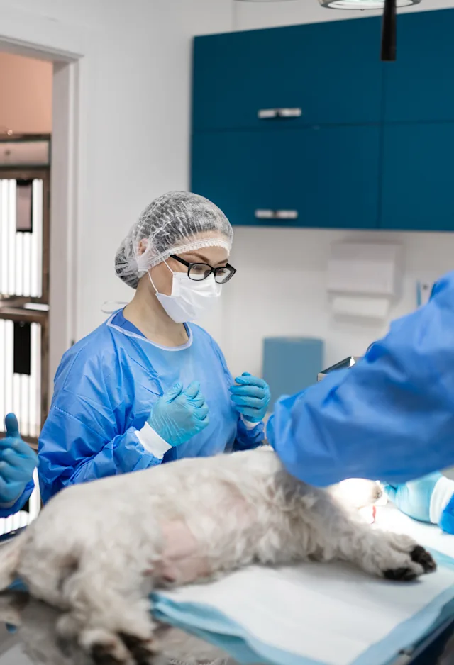
A veterinary cardiologist is a specialized veterinarian with advanced training and expertise in the diagnosis, treatment and prevention of heart and vascular diseases. Veterinary cardiologists are also knowledgeable about pulmonary (lung) diseases.
A board certified veterinary cardiologist is a veterinarian who has obtained intensive, additional training in cardiology and has been certified by the American College of Veterinary Internal Medicine (DACVIM - Cardiology). The advanced training and education required for this certification includes:
- A veterinary degree (three to four years of undergraduate college, followed by four years of veterinary school).
- One year rotating internship in veterinary medicine and surgery.
- Three years of advanced cardiology training (residency), including extensive training in a variety of diagnostic imaging techniques (radiology, echocardiography and angiography) and interventional cardiac procedures.
- A series of rigorous examinations covering all aspects of general internal medicine and veterinary cardiology.
Philosophy:
When your pet needs the care of a veterinary cardiologist, years of intensive training and education will be focused on helping your pet to enjoy the best quality of life possible. The Cardiology service at CVRC aims to improve the management and long term prognosis of any animal with heart disease. We will work closely with your primary care veterinarian so that your pet’s care will be shared by the primary care doctor who knows him/her the best and a heart specialist. Including a cardiologist in your pet’s care if they have heart disease will improve their outcome and quality of life. Appropriate treatment can often prolong the time before symptoms of heart disease develop. In dogs and cats with symptoms of heart disease, treatment can reverse the symptoms and allow your pet additional months to years of a comfortable and happy life.
What is an emergency/specialty hospital and how is it different than my primary veterinarian?
A specialty hospital does not offer any routine or preventative care. Our veterinarians have advanced training in specific disciplines such as surgery, oncology, internal medicine, emergency and critical care medicine, physical rehabilitation and neurology among others. We also have equipment that most primary veterinarians don’t have such as CT scans, MRI, endoscopic equipment, and specialized surgical tools. We work closely with your primary veterinarian to offer these services to you.
What diagnostics/procedures are offered?
Physical examination: Physical exam is the cornerstone of every good workup. A full physical exam focused on the heart and lungs will be performed by the cardiologist at CVRC during your pet’s appointment. Physical exam abnormalities (heart murmur, arrhythmia, trouble breathing, e.g.) will help define which diagnostic tests are the most appropriate for your pet.
Cardiac Imaging:
- Echocardiography (Echo): Echocardiography is an ultrasound of the heart which shows its structure and function. An echo is by far the best way to assess heart disease, because it shows things we cannot see on chest X-rays (better assessment of chamber size, contractility, valve shape, etc.) or hear during osculation (listening with stethoscope). An echo will be recommended in most patients on their first visit to CVRC as this is the best way to make a specific diagnosis, identify severity of disease, and formulate a monitoring and treatment plan. Echo's are non-invasive and not dangerous. An echo requires specialized ultrasound equipment and ideally should be performed by someone with advanced education and training, preferably a board certified cardiologist. At CVRC, we generally perform echo's without sedation and they are painless.
- Doppler echocardiography: Doppler echo is an advanced form of echocardiography in which the direction and speed of blood flow in the heart and great vessels can be visualized and documented. It is essential in quantifying the severity of some forms of heart disease, in assessing for high blood pressure within the lungs (pulmonary hypertension), and in assessing the risk of heart failure (fluid in the lungs) occurring. Doppler echo provides significantly more information about heart disease than standard echocardiography, and is part of every echo performed at CVRC.
- Chest X-Rays: Chest X-rays are taken to evaluate the heart and lungs. At CVRC, all X-rays are digital and of the highest quality. Interpretation by board certified radiologists is available when indicated. Chest X-rays might be recommended in a patient who is having trouble breathing or exhibiting a cough. Chest X-rays are necessary to make a diagnosis of heart failure (fluid in the lungs).
ECG/EKG
- 6-Lead Electrocardiogram (ECG or EKG): This is the test that shows electrical function of the heart. An ECG is necessary for any animal diagnosed with an arrhythmia, or irregular heart rate or rhythm. A 6-lead ECG provides more information than a single lead ECG, and all ECGs performed by the Cardiology service at CVRC are 6-lead. The ECG allows for a specific arrhythmia diagnosis which is necessary to gauge the risk of the arrhythmia and to formulate a treatment plan.
- Holter Monitor (ambulatory ECG monitoring): A Holter is an ambulatory ECG monitor that your pet would wear home in a little backpack. CVRC’s digital Holter units can record heart rate and rhythm for up to 48 hours (although 24 hours is standard) and results are usually back in a few days. Holters increase the ECG sampling period tremendously compared with an ECG performed during an appointment, so it will be recommended in animals in which the in-house 6-lead ECG does not provide enough information about the significance of an arrhythmia.
Blood Pressure Monitoring: Blood pressure is affected by heart disease and high blood pressure can increase the work of the heart. For this reason, if your veterinarian hasn’t done so recently, blood pressure is often checked when your pet has an appointment with the Cardiology service at CVRC. The technique is non-invasive, nonpainful, and involves an inflatable cuff placed around a limb or tail and a special monitor.
Bloodwork: Some heart medications require bloodwork monitoring to ensure they are working properly and not having systemic side effects. If your veterinarian hasn’t performed bloodwork recently, it may be recommended when you bring your pet to the Cardiology service at CVRC. Some bloodwork, such as checking electrolytes and kidney values, is performed in-house so results will be available during your pet’s appointment. Other bloodwork might be sent out to other laboratories, in which case we will contact you when results return.
Fluid Taps: Heart disease can sometimes lead to the accumulation of fluid in the chest or abdominal cavity or the pericardium (sac surrounding the heart). This fluid accumulation can be life threatening and might sometimes require a removal, which can be performed at CVRC. This is a procedure in which a needle is inserted into the chest cavity, abdominal cavity, or pericardium and fluid is removed. Your pet might be lightly sedated for this procedure for his or her safety.
Angiography: Angiography refers to delivering contrast or dye through an intravenous catheter into the bloodstream so that blood flow can be seen using fluoroscopy (real time continuous X-ray). Angiography is performed at CVRC in the Cath Lab to better identify heart and vessel size and shape in order to guide interventional procedures such as patent ductus arteriosus (PDA) closure and balloon dilation valvuloplasty for pulmonic stenosis (PS) and subaortic stenosis (SAS).
Cath Lab and Interventional Procedures: Using extensive specialized equipment, training, and expertise, minimally invasive procedures to correct or address heart or blood vessel abnormalities can be performed. These procedures require the use of fluoroscopy and include pacemaker placement, patent ductus arteriosus (PDA) closure and balloon dilation valvuloplasty for pulmonic stenosis (PS) or subaortic stenosis (SAS).
Pacemaker Placement: Pacemaker placement is the most common interventional Cath Lab procedure that we perform. It involves accessing the heart via a peripheral blood vessel, and implanting a set of electrodes (called leads) into the heart to correct for certain arrhythmias. The lead is connected to the pacemaker device, which sends the electrical impulses through the leads to the heart. The pacemaker device is implanted under the muscle on the side of the neck, while the lead extends through the jugular vein into the heart. We have the ability to adjust the programming of the pacemaker onsite when needed.
Oxygen Therapy: Oxygen supplementation can be delivered to your pet when indicated via flow by, intranasal, or environmentally controlled oxygen cage. Delivering a higher percentage of oxygen to the lungs can help maximize oxygen delivery to tissue in cases of respiratory or cardiac dysfunction.
What should I expect during my visit?
If you are a new client to CVRC, we encourage you to fill out our New Patient Registration Form ahead of time online, or bring the form with you. Otherwise, please arrive to your appointment approximately 10 minutes early to complete this form in our office.
Visits with the Cardiology service at CVRC tend to take about an hour. If your pet’s care is more complicated, additional time might be needed. Your visit will start with the cardiology technician meeting you and your pet and getting a full history from you. Please bring any medications that your pet is taking so that we can confirm dosing. The cardiology technician or the cardiologist will go over diagnostic plans and costs with you. After making a plan for diagnostics, we will take your pet to our treatment area or echocardiography suite to perform any tests (echocardiogram, ECG, X-rays, blood pressure, bloodwork, etc.). When testing is done, we will return your pet to you while the cardiologist is interpreting tests and writing a summary report. The cardiologist will explain the results of the exam and diagnostic tests after they are completed and answer any questions you may have. We will also communicate with your primary veterinarian during this process to ensure a collaborative treatment plan for the care of your pet. You will be given a printed visit summary with the diagnosis, treatment recommendations, and follow up care. A full report will be sent to your family veterinarian as well. We welcome your progress reports, questions and concerns any time and will maintain an ongoing relationship with your primary care doctor to ensure the best experience for you and the best care for your pet.
What should I bring to my visit?
First and most importantly, bring your pet to all visits. Follow your normal routine for medications and feeding the day of your visit, unless you have been instructed otherwise by our staff. Also, please bring all medications that your pet is receiving so that we can verify the dosages. Ensure that the person bringing your pet in has a good knowledge of your pet's medical history, as this is an important part of the consultation. Records can be faxed or emailed to us from your primary care veterinarian prior to the appointment. If your pet has had any x-rays taken recently, please bring those with you as well. We can request records and digital x-rays on your behalf from your family veterinarian once you have scheduled a visit with us. Ensuring that you bring all related information will minimize the risk of needed to repeat any diagnostics or tests.
What is echocardiography?
Echocardiography is a type of ultrasound that uses special probes and software, as well as requiring advanced training, to perform real time imaging of the heart and surrounding structures. At CVRC, echocardiograms are performed by our board certified specialists, who have undergone extensive education and training in echocardiography to aid diagnosis and treatment of all cardiac-related diseases.
How is echocardiography performed?
After being thoroughly examined by one of our doctors, your pet is positioned appropriately on the echocardiography examination table. For most patients, sedation or anesthesia is not required. The area of interest is clipped to allow the probe to make contact with the skin. The study is performed and interpreted by one of our trained clinicians. The procedure takes approximately 30 minutes to perform. Echocardiography is painless and noninvasive.
What is a heart murmur?
A heart murmur is an abnormal sound heard on physical exam with a stethoscope, which develops when blood flow in the heart or great vessels is turbulent. This occurs when blood flow is too fast and is often (although not always) a sign of heart disease. The loudness of a heart murmur (grade) is related to the severity of the heart disease in some cases, but not all. If the disease is severe, your pet might be at risk to show symptoms of heart disease (trouble breathing, cough, exercise intolerance, lethargy, abdominal distention, weakness, collapse, etc.). The best way to investigate the cause and significance of a heart murmur is with heart imaging, and echocardiography is superior to chest X-rays for this purpose. Echo's and chest X-rays are offered at CVRC through the Cardiology service. Information provided by echo will be used to formulate the best treatment plan to keep you pet as comfortable and happy as possible for as long as possible.
What does it mean if my puppy or kitten has a heart murmur?
Heart murmurs can be benign, significant, or somewhere between. If your puppy or kitten has a loud heart murmur, it is probably because he or she was born with a heart defect. An echocardiogram (echo) is strongly recommended for these cases, as some heart defects are fixable in the Cath Lab, while others can be managed with medications to improve quality of life and life expectancy. A workup should be performed sooner rather than later, so that management can be started before the heart changes irreversibly and symptoms develop. Waiting to perform a workup until clinical signs develop worsens the prognosis. If your puppy or kitten has a soft heart murmur, it might still be because he or she was born with a defect, but a soft heart murmur can also be completely benign (not associated with underlying disease). An echo is the only way to know the difference. If you are going to have an echo performed in a young animal with a heart murmur, it should be a complete echo with Doppler performed by a cardiologist. Heart defects can be very complicated and cardiologists are specially educated and trained in this area. Doppler provides information about severity of disease, which is essential to guide treatment plans.
If my pet was born with a heart defect, can it be fixed?
Some heart defects that animals are born with can be completely fixed, while others can be improved by interventional procedures performed in the Cath Lab. These procedures are modeled after interventional procedures performed in people. We can fix or improve some heart defects by accessing the heart through catheters placed in peripheral veins and arteries in the neck and the groin. In this way, we can treat heart defects that used to be treatable only with open chest surgery (sometimes open heart surgery). Other heart defects that your pet might have been born with can be better managed with medications. After the workup at CVRC, the cardiologist will speak with you about what the options for treatment are.
What is an arrhythmia?
An arrhythmia is an abnormal heart rate or rhythm. Arrhythmias are a consequence of dysfunction of the heart’s electrical system. An ECG is required to diagnose what arrhythmia your pet has, which is necessary to make the best treatment and monitoring plan. If severe, arrhythmias can cause symptoms such as weakness, collapse, restlessness, and even sudden death. Treatment for arrhythmias depends on whether the arrhythmia is too fast or too slow. If the arrhythmia is too fast, treatment is usually with medications. If the arrhythmia is too slow, treatment is usually pacemaker implantation. The cardiologist at CVRC will be able to interpret the ECG and explain the significance, treatment options, and prognosis with you.
What is heart failure?
Heart failure is a group of symptoms that occurs when the heart can no longer keep blood moving forward effectively. It results in the accumulation of fluid in or around the lungs (or less commonly, in the abdomen). The most common and significant symptom of heart failure is trouble breathing. Other symptoms include cough, exercise intolerance, lethargy, decreased appetite and abdominal distention. Chest X-rays are required to diagnose most cases of heart failure. If your pet has heart failure diagnosed, a workup with the cardiologist at CVRC will help optimize the treatment and monitoring plan for your pet, increasing life expectancy for as long as possible. Animals can be effectively treated for heart failure for over a year, with a good quality of life. If your pet is diagnosed with heart failure at your primary veterinarian's clinic and needs to be provided with 24 hour care or more extensive treatment and monitoring, CVRC can provide that so your pet can be feeling better as soon as possible to be able to go home.
What does a large heart on chest X-rays mean?
A large heart on chest X-rays usually signifies heart disease of some variety. If your veterinarian is concerned that your pet has a large heart on chest X-rays, an echocardiogram (echo) would be the next diagnostic step. An echo is much more sensitive and gives more information than a chest X-ray to identify specific heart diseases (many of which can cause a large heart), and is needed in order to optimize management and outcome. Sometimes, an echo will show that the heart is actually normal, even though it has looked abnormal on chest X-rays. This also is important to know so that unnecessary medications or treatments can be avoided.
What is the Cath Lab and what is an interventional procedure?
The Cath Lab is the room in which the cardiologist at CVRC performs interventional procedures. These procedures require extensive specialized equipment and training. They are minimally invasive procedures in which the heart is accessed using catheters placed in the peripheral blood vessels in the neck and groin. In this way, heart disease can be treated without open chest or open heart surgery. The most common cardiac procedure performed in the Cath Lab is pacemaker implantation in dogs that are collapsing because of very slow heart rates. Other procedures include patent ductus arteriosus (PDA) closure and balloon dilation valvuloplasty for pulmonic stenosis (PS), PDA occlusion, balloon valvuloplasty OR stenting for pulmonic stenosis, and heartworm extractions for life-threatening heartworm disease complications. These procedures can cure or improve heart diseases, leading to a better quality of life and life expectancy.
Do I need a referral?
While referrals are recommended to see one of our specialists, they are not required. It is always better to speak to your veterinarian about a referral so we can collaborate with your veterinarian to ensure the best care for your pet.
Will you keep my family veterinarian informed of the care my pet receives at CVRC?
We work closely with your primary veterinarian and ensure they receive copies of all medical records. We also communicate via phone and they have the ability to access a portal into our medical record system.