

Imaging Overview
High quality digital imaging equipment is an essential and integral component for diagnosis and treatment of our patients. CVRC has invested in comprehensive, complementary, and high quality imaging modalities to support our clinicians’ ability to visualize the internal body structures of our patients. Diagnostic imaging often requires the use of several of these different technologies to obtain the information needed to diagnose and treat our patients. These imaging modalities include digital radiography, ultrasound, echocardiography, CT Scan, C-arm (fluoroscopy), MRI, arthroscopy, laparoscopy, thoracoscopy, and flexible and rigid endoscopy.
We can easily share patient’s images with our staff clinicians, the primary veterinarian, and other specialists across the country (through telemedicine) for prompt review, interpretation, and collaboration, ensuring the highest level of care for our pet patients.
Digital X-ray (DR)
X-ray imaging is used to visualize internal structures of patients, and is used for a wide range of applications including imaging of the thorax, abdomen, skeletal structures, limbs and a variety of special imaging procedures. DR images are available within seconds to be viewed on a high-resolution monitor with no need for film developing. Examination rooms are equipped with flat screen LCD viewing stations for reviewing the images with pet owners.
All radiographic studies are interpreted by board certified specialists.
What are the advantages of Digital X-ray vs traditional film X-ray?
- Superior image quality.
- Faster study times (Approximately 4 seconds to acquire each image)
- More efficient – more X-ray studies can be performed in less time
- Less anxiety for the animal from reduction in study time
- Reduces patient and staff exposure time to radiation
- Allows for superior image manipulation
- Instantaneous viewing on all computers in the hospital
- All images can be viewed and assessed by a Board Certified Veterinary Radiologist
via telemedicine
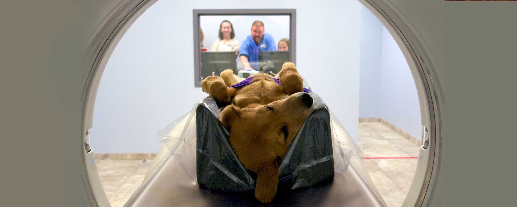
CT Scan
CT scan is a non-invasive imaging modality that performs 360◦, cross-sectional imaging of our patients. CT scan uses X-ray technology to acquire these images, but does so with vastly increased detail and sensitivity, allowing us to visualize structures less than 1mm in size.
Our 16 slice, GE Optima 520 CT scanner is able to perform imaging studies much faster than older, conventional scanners that are often found in veterinary medicine. Using newer equipment and software, we are able to perform most studies in less than a minute (actual scanning time). This reduction in time also comes with increased quality and sensitivity: advantages that translate to shorter anesthetic times for our patients, even the ability to perform some studies under a light plane of sedation.
When more complex issues arise with your pet's health, our doctors look to our state-of-the-art diagnostic imaging technology, including on-site CT, to provide information that otherwise cannot be found by physical examination or bloodwork alone.
How is CT performed?
After being thoroughly examined by one of our doctors, your pet is sedated or anesthetized and placed on the CT imaging table. The CT technician positions the patient and programs the CT computer for the desired type of study. The patient table then advances through the CT gantry (which is a large, donut shaped opening) while an X-ray tube and sensors rotate at high speed around the patient. Usually, the scan is performed twice, the second time with administration of an intravenous contrast agent that highlights blood vessels and other structures. These images are reconstructed on a computer console attached to the CT machine, then reviewed by our specialists.
What is CT used for?
Brain – Trauma with suspected fracture, acute intracranial injury in an unstable patient
Spine – Fractures, mineralized disc rupture, neoplasia
Nasal Cavity & Sinuses – Chronic nasal discharge and/or sneezing, nasal distortion/deformation, neoplasia
Orbit/Ocular – Orbital trauma with suspected fracture, neoplasia
Head/Neck – Dental-related neoplasia, head/neck trauma with suspected fracture
Thorax – Metastatic screening, primary lung neoplasia, pneumothorax, lung lobe consolidation, chronic pneumonia, pleural effusion, mediastinal disease
Abdomen – Upper urinary tract evaluation, ectopic ureter, renal/ureteral calculi, clarification and surgical planning for large organ masses (including liver, spleen, kidney, GI tract), investigation of suspected portosystemic shunts
Musculoskeletal – Elbow dysplasia, suspected incomplete ossification of the humeral condyles, complex fractures
Neoplasia – Surgical planning for tumor removal or debulking, lung metastasis screening
Cardiovascular – Pulmonary embolism
Other – General trauma
**CT and MRI overlap in their use, and the decision of one modality over another is based on availability, cost, anticipated findings and patient stability.**
MRI
Magnetic Resonance Imaging (MRI) is an advanced, non-invasive, non-radiating imaging modality that has become commonplace in human medicine, and has rapidly expanded into the veterinary field.
MRI uses a strong magnetic field that aligns protons of the body tissues within the field. A radio frequency signal (RF) is then applied to the field which disrupts the alignment of these protons, after which they return to their original state. The RF signals are collected by a receiver (called a coil) and then transmitted back to a computer, which creates visual images from this information.
These images are easily constructed in axial, dorsal, sagittal and oblique planes for review by the clinician or radiologist. As in CT, MRI is often performed using an intravenous contrast agent that enables us to better visualize certain structures and abnormalities. MRI yields the highest level of soft tissue detail compared to other imaging modalities (CT, digital X-ray). This sensitivity allows for highlighting desired structures or tissues by adjusting settings on the MRI machine.
How is an MRI performed?
After being thoroughly examined by one of our doctors, your pet is anesthetized and placed in the MRI. The MRI technician positions the patient and programs the MRI computer for the desired type of study. An MRI study typically takes 30 minutes to one hour to complete, and requires general anesthesia, as the animal must be perfectly still for the duration of the study. The images are reconstructed on a computer console attached to the MRI, then reviewed by our specialists.
MRIs are performed by technicians who have received advanced education and training in magnetic resonance imaging.
What is MRI used for?
Brain – Brain disorders, seizures, cranial nerve signs, vestibular disease, ataxia, head tilt, behavioral changes, central blindness, pituitary disease, metastatic disease, congenital abnormalities, head trauma
Spine – Acute or progressive tetraparesis or hindlimb paresis, CP deficits, spinal pain, nerve root signature, spinal trauma, congenital abnormalities, discospondylitis
Peripheral Nervous System – Suspected peripheral nerve neoplasia, progressive single limb atrophy or pain
Nasal Cavity & Sinuses – Chronic nasal discharge and/or sneezing, nasal distortion/deformation, neoplasia
Orbit/Ocular – Exophthalmous, pain opening mouth, retrobulbar swelling or neoplasia, optic nerve disease
Head/Neck – Soft tissue masses or swelling, including lymph nodes, salivary glands, larynx, thyroid, maxilla, mandible, chronic ear disease, head tilt
Abdomen – Small organ masses including the pancreas, lymph nodes and adrenal glands, surgical planning for clarification or margins and involvement of neoplasia of large organs, including the liver, spleen, kidney, and GI tract
Musculoskeletal – Pain or lameness localized to a joint in the limb (particularly ligaments, tendons, menisci, cartilage, bone, or joint swelling)
Neoplasia – Soft tissue masses, especially for surgical planning, metastatic screening, lymph node and bone marrow evaluation
Cardiovascular – Pericardial effusions, pericardial masses, suspected large vessel thrombus
Other – Suspected foreign bodies, chronic draining tracts
**CT and MRI overlap in their use, and the decision of one modality over another is based on availability, cost, anticipated findings and patient stability.**
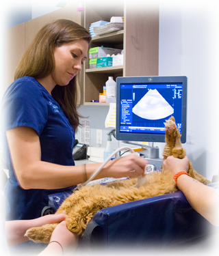
Ultrasound
Ultrasound is used as a diagnostic tool to obtain more detailed information about internal body structures. Ultrasound machines use sound waves administered by a small handheld device called a probe, waves are reflected back to the probe, and a computer formats these into visual pictures on a monitor for the clinician to review. Ultrasound is painless and noninvasive.
Ultrasound is a modality that requires significant training and experience by the clinician to achieve an accurate diagnosis and interpretation of the study.
How is ultrasound performed?
After being thoroughly examined by one of our doctors, your pet is positioned appropriately on the ultrasound examination table. For most patients, sedation or anesthesia is not required. The area of interest is clipped to allow the probe to make contact with the skin. The study is performed and interpreted by one of our trained clinicians. The procedure takes approximately 30 minutes to perform. Ultrasound is painless and noninvasive.
What is ultrasound used for?
Applications for ultrasound include:
- Evaluation of the abdominal organs, including the stomach, liver, spleen, pancreas, gastrointestinal tract, lymphatic system, kidneys, urinary tract, and endocrine organs
- Pregnancy evaluation
- Imaging of thoracic and abdominal masses
- Imaging of fluid accumulation in the abdomen or thorax
- Imaging of the neck, including thyroid and parathyroid glands
- Imaging of blood vessels and flow, in and around the heart, and throughout the body
- Imaging of the eye (for masses and retinal detachment)
- Assessment of internal injuries after trauma (called an AFAST/TFAST scan)
- Minimally invasive techniques to obtain samples of organs for diagnosis of illnesses and cancers (fine needle aspirates, ultrasound-guided biopsies and cystocentesis, gall bladder secretions)
Echocardiography
Echocardiography is a type of ultrasound that uses special probes and software, as well as requiring advanced training, to perform real time imaging of the heart and surrounding structures. At CVRC, echocardiograms are performed by our board certified specialists, who have undergone extensive education and training in echocardiography to aid diagnosis and treatment of all cardiac-related diseases.
How is echocardiography performed?
After being thoroughly examined by one of our doctors, your pet is positioned appropriately on the echocardiography examination table. For most patients, sedation or anesthesia is not required. The area of interest is clipped to allow the probe to make contact with the skin. The study is performed and interpreted by one of our trained clinicians. The procedure takes approximately 30 minutes to perform. Echocardiography is painless and noninvasive.
What is echocardiography used for?
- Pericardial effusions
- Dilated cardiomyopathy
- Mitral regurgitation
- Hypertrophic cardiomyopathy
- Pulmonary stenosis
- Ventricular septal defects
- AV valve dysplasia
- Patent ductus arteriousus
- Developmental defects
- Pulmonary hypertension
- Heartworm disease
- Heart based masses
- Advanced feline restrictive cardiomyopathy
- Right to left shunts
- Atrial septal defects
- AV valve stenoses
- Coronary artery defects
- Occult dilated cardiomyopathy
- Cor triatriatum
- Persistent left cranial vena cava
- Canine hypertrophic cardiomyopathy
- Feline diastolic dysfunction
Endoscopy
Endoscopy is a minimally invasive technique that uses flexible and rigid scopes to visualize internal structures of a patient, including the upper and lower gastrointestinal tract, nasal passages, respiratory tract, and urinary tract.
How is endoscopy performed?
After a full physical examination, the patient is sedated or anesthetized. A specialized scope is passed into the area of interest (gastrointestinal, urinary, or respiratory tract), and a camera hooked up to the scope displays live images of these structures. Using special instruments, biopsy samples can be obtained, as well as foreign material removed.
What is endoscopy used for?
Endoscopy is used to visualize the gastrointestinal, respiratory and urinary tract, and to obtain diagnostic tissue samples to diagnose conditions affecting these body systems. Polyps can be removed. Endoscopy can also be used to extract upper gastrointestinal foreign material, and to assist in placement of feeding tubes.
Arthroscopy - Laparoscopy - Thoracoscopy
Arthroscopy involves the passage of a rigid scope into a joint, connected to a camera and monitor, to visualize the inside of a joint and certain procedures can be performed to help diagnose and treat various conditions, as well as obtain diagnostic samples for laboratory analysis.
Laparoscopy involves the passage of a rigid scope into the abdominal cavity, connected to a camera and monitor, to visualize abdominal organs, and certain procedures can be performed to help diagnose and treat various conditions, as well as obtain diagnostic samples for laboratory analysis.
Thoracoscopy involves the passage of a rigid scope into the thoracic cavity, connected to a camera and monitor, to visualize thoracic organs, and certain procedures can be performed to help diagnose and treat various conditions, as well as obtain diagnostic samples for laboratory analysis.
All three of these procedures require anesthesia and are performed in a sterile operating room suite by one of our board certified specialists. Additional information on these procedures is available on our surgical services page.
Fluoroscopy
Fluoroscopy is an imaging technique that uses X-rays to obtain real-time moving images of the internal structures of a patient. Fluoroscopy is used for dynamic studies to look for functional abnormalities in various body systems. Additionally, fluoroscopy is used to guide implant placement or to help obtain tissue samples. Depending on the type of study, sedation or anesthesia may be necessary.